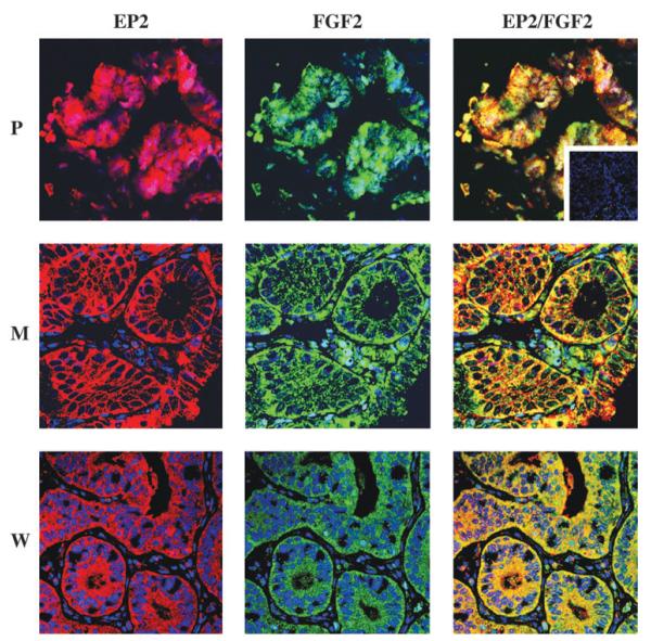
Figure 5.
Confocal immunofluorescent localization of E-series prostanoid-2 (EP2) receptor and fibroblast growth factor 2 (FGF2) in endometrial adenocarcinoma. Localization of the site of expression of the EP2 receptor (red; left panel), FGF2 (green; central panel) and co-localization of EP2 receptor and FGF2 (merged yellow; right panel) in human endometrial adenocarcinoma. P, poorly differentiated; M, moderately differentiated and W, well-differentiated adenocarcinomas; inset, negative control section.

