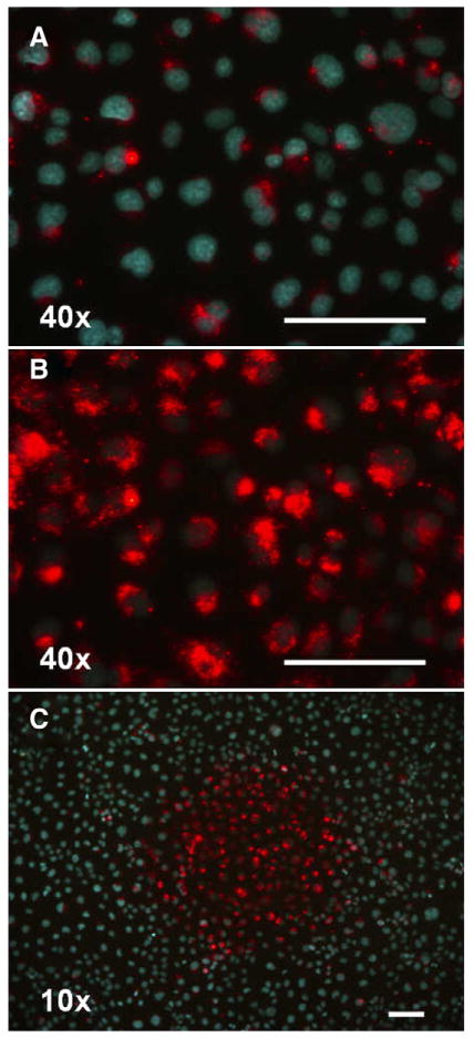
Fig. 4.
Enhancement of fluorescence of quantum dots (8 nM QD655-carboxyl terminated) endocytosed in Du145 cells (5×10⁵ cells/ml). A) Fluorescence microscopy photograph (objective 40×) taken within 0–1 min of observation after around 24 h incubation. Nuclei are stained with Hoechst 33342. B) The same spot on the culture dish photographed under the same conditions after 5 min irradiation with the microscope excitation light (395–440 nm). Red fluorescence appears while the Hoechst dye is photobleached during irradiation. C) Fluorescence photograph taken at lower magnification (10×) highlights the red fluorescent spot caused by the irradiation using the 40× objective. The scale bars are 100 μm.

