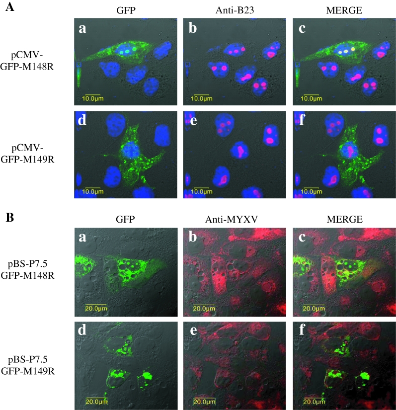
Figure 3.
Cellular localization of M148R and M149R. (A) BGMK cells grown on coverslips were transfected with pCMV-GFPM148R (a, b, c) or pCMV-GFPM149R (d, e, f). Cells were fixed at 24 h p.t., permeabilized, stained with anti-B23 antibody (red staining) and TO-PRO-3 iodide to detect nuclei (blue staining), and analyzed by confocal microscopy with a 60× objective. Multistaining experiments were performed using the sequential mode. (B) BGMK cells grown on coverslips were pBS-P7.5GFPM148R (a, b, c) or pBS-P7.5GFPM149R (d, e, f) transfected, with prior 2 h infection with the MYXV deleted for the corresponding gene. At 24 h p.t., cells were fixed, permeabilized, stained with anti-MYXV serum (red staining) and analyzed by confocal microscopy as previously indicated.

