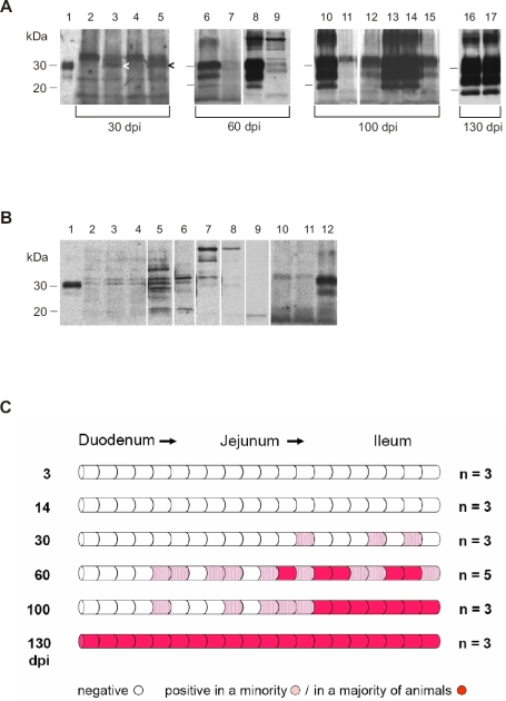
Figure 3.
(A) Western blot detection of PrP27-30, the PK-resistant core of PrPTSE, in segments of the small intestine from hamsters fed with 263K scrapie brain homogenate. Lane 1: Blot control, 263K scrapie hamster brain homogenate containing 1 × 10⁻⁷ g of brain tissue. Lanes 2 to 5: Samples collected at 30 dpi (weak specific immunostaining signals for PrPTSE in lanes 3 and 5 are marked by arrowheads). Lanes 6 to 9: Samples collected at 60 dpi (adjacent intestinal samples were blotted in lanes 6 and 7, and in lanes 8 and 9). Lanes 10 to 15: Samples collected at 100 dpi (adjacent intestinal samples were blotted in lanes 10 and 11, and in lanes 12–15; the pattern shown in lanes 12–15 would be compatible with bi-directional spread of infectivity in the wall of the intestinal tract. Lanes 16 and 17: Samples collected 130 dpi. Note: Positive samples at 100 and 130 dpi also show strong staining of PrP27-30 dimers. (B) Range of western-blot signals from different segments of small intestine from uninfected hamsters after PrPTSE purification and staining with mAb 3F4. Lane 1: Blot control, 263K scrapie hamster brain homogenate containing 1 × 10⁻⁷ g of brain tissue. Lanes 2–11: Unspecific immunostaining in extracts from different segments of the small intestine of uninfected hamsters. Lane 12: Positive control; tissue sample collected at 60 dpi from the small intestine of a hamster orally infected with 263K scrapie. (C) Initial sites, foci, and spread of PrPTSE-deposition in the small intestine of hamsters fed with 263K scrapie brain homogenate. Schematic summary of Western blot findings in screened segments of small intestines collected 3, 14, 30, 60, 100 and 130 dpi. n = number of animals investigated.

