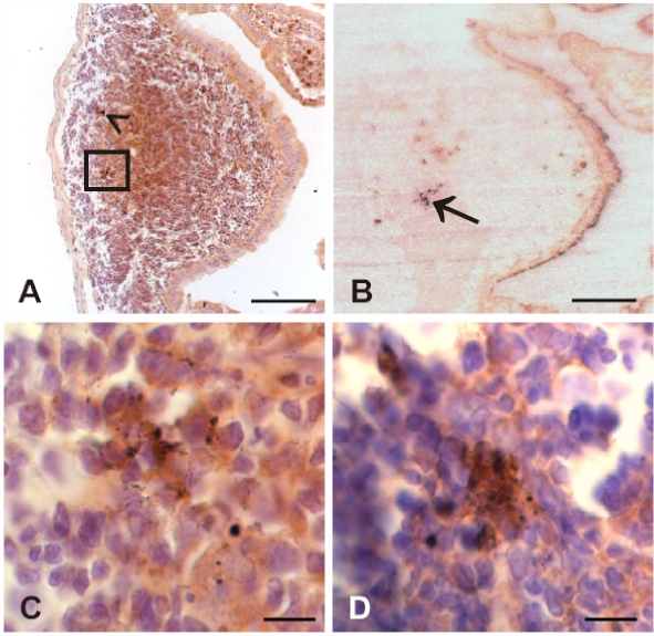
Figure 4.
Photomicrographs displaying granularly stained PrPTSE detected by immunohistochemical labeling (A, C, D) or PET blotting (B) at 30 or 45 dpi in the GALT of hamsters fed with 263K scrapie brain homogenate. PrPTSE appears as brown (A, C, D) or blue-magenta (B) granular material. (A) Ileal lymphoid follicle at 45 dpi showing PrPTSE in the germinal centre (arrowhead and inset in A). (B) PET blot in adjacent section to the specimen displayed in A (PrPTSE is indicated by arrow). (C) Higher magnification of inset in A. (D) Detection of PrPTSE in the germinal center of an ileal lymphoid follicle at 30 dpi. Scale bars in A and B, 40 μm; in C and D, 5 μm. (For a colour version of this figure, please consult www.vetres.org.)

