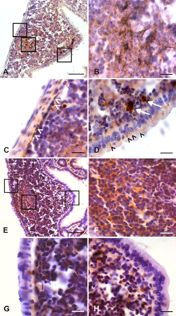
Figure 5.
Photomicrographs displaying PrPTSE detected by immunohistochemical staining at 60 dpi in the GALT of a hamster fed with 263K scrapie brain homogenate (A–D). Lymphoid follicle from the distal jejunum showing granular deposits of PrPTSE in the germinal center (A, middle inset; B, higher magnification of middle inset in A), in submucosal nerve-like structures (A, left inset; C, higher magnification of left inset in A, white arrowheads), in cells of the FAE and in macrophages of the dome (A, right inset; D, higher magnification of right inset in A, black arrowheads and white arrows, respectively). Scale bars in A, 40 μm; in B, C and D, 5 μm. Photomicrographs of a lymphoid follicle from the distal ileum of an uninfected hamster labeled immunohistochemically with mAb 3F4 (E–G). Unspecific homogeneous brown staining in the germinal center (E, middle inset; F, higher magnification of middle inset in E). Granular immunostaining for PrPSc deposits is absent in the submucosal region (E, left inset; G, higher magnification of left inset in E), or in the FAE (E, right inset; H, higher magnification of right inset in E). Scale bars in E, 40 μm; in F, G and H, 5 μm. (For a colour version of this figure, please consult www.vetres.org.)

