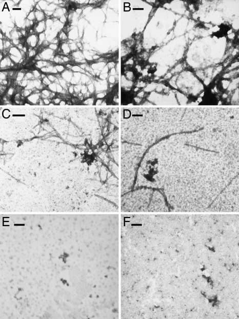
Fig. 4.
Ligands affect fibril formation of Aβ1–40. Transmission electron microscopy show fibrils formed from (A) 25 μM Aβ1–40 incubated alone or with addition of 25 μM of (B) Pep1a, (C) Pep1b, or (D) Dec-DETA. The bottom row shows incubated samples containing (E) 125 μM Pep1b and (F) 125 μM Dec-DETA alone. All incubations were done at 37 °C with agitation. (Scale bars, 100 nm.)

