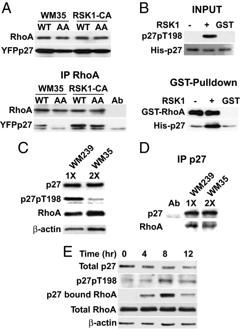
Fig. 5.
p27 T198 phosphorylation increases RhoA binding. (A) WM35 and RSK1-CA were transfected with YFP-p27WT (WT) or YFP-p27T157A/T198A (AA). Cellular RhoA and YFP-p27 (Upper) and RhoA-bound YFPp27 (Lower) are shown. (B) His-p27 was treated (+) or not (−) with RSK1 in vitro, then recovered and incubated with GST-RhoA or control (GST). RhoA-bound His-p27 was detected by GST pull-down. His-p27 and p27pT198 inputs are shown. (C and D) Cellular p27 was titrated in WM35 and WM239 to allow precipitation of equal amounts of p27. p27 in WM35 was roughly half that in WM239. (C) Western blots show equal p27 in 10 μg of WM239 lysate (1X) and 20 μg of WM35 lysate (2X). Input p27pT198, RhoA, and β-actin levels are shown. (D) Equal p27 amounts were precipitated and resolved, and associated RhoA was blotted. (E) WM35 cells were synchronized in G0 and released into cycle at 0 h. At 4-h intervals thereafter, total p27, p27pT198, RhoA, and β-actin were blotted and p27-bound RhoA was detected.

