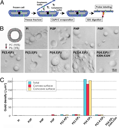
Fig. 1.
Labeling of the liposome. (A) Outline of the method. Cells were rapidly frozen, freeze-fractured, and evaporated with carbon (C) and platinum/carbon (Pt/C) in vacuum. The replica of the split membrane was digested with SDS to remove noncast molecules and labeled by GST-PH. Both the cytoplasmic and exoplasmic halves of the membrane were examined. (B) Labeling of small unilamellar liposome replicas. Freeze-fracture replicas of liposomes containing 95 mol % of phosphatidylcholine (PC) and 5 mol % of phosphatidylinositol or a phosphoinositide were labeled. Only liposomes containing PI(4,5)P2 were labeled intensely by GST-PH. A PH mutant, GST-PH(K30N, K32N), which does not bind PI(4,5)P2, showed little labeling in the PI(4,5)P2-containing liposome. (C) Quantification of the GST-PH labeling in the liposomes. The number of gold particles per 1 μm2 of the liposome surface is shown (blue). The labeling on the convex (red) and concave (yellow) surfaces showed equivalent results.

