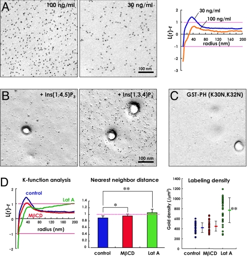
Fig. 2.
Labeling in the flat undifferentiated area of the cell membrane. (A) Distribution of PI(4,5)P2 in the P face of the flat undifferentiated cell membrane of the human fibroblast in culture. GST-PH was used at a concentration of 100 ng/mL (Left) or 30 ng/mL (Right). The mean L(r) − r curve compiled from 30 randomly chosen areas showed that the labeling by 100 ng/mL GST-PH was randomly distributed, whereas that by 30 ng/mL GST-PH showed weak clustering. (B) Effects of inositol trisphosphates on the PI(4,5)P2 labeling. GST-PH (100 ng/mL) was preincubated with either 1 mM Ins(1,4,5)P3 or 1 mM Ins(1,3,4)P3 before applying to replicas. Ins(1,4,5)P3 abolished the labeling completely, whereas Ins(1,3,4)P3 did not. (C) Labeling by GST-PH (K30N, K32N) used at 100 ng/mL. Virtually no labeling was observed. (D) The clustering that was detected by 30 ng/mL GST-PH decreased when cells were treated with 5 mM methyl-β-cyclodextrin (MβCD) for 60 min to extract free cholesterol or with 1 μM latrunculin A (Lat A) for 10 min to depolymerize actin. After these treatments, the normalized nearest neighbor distance became closer to one, and the average labeling density increased (*, P < 0.01; **, P < 0.001).

