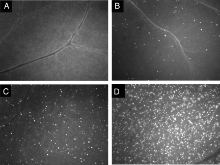
Fig. 3.
Higher magnification images of retinal flatmounts from rat eyes treated with intravitreal NMDA and subsequently injected with TcapQ. Fluorescence microscopic photographs of representative retinal flatmounts from PBS-pretreated eyes (A), and NMDA-pretreated eyes with injectate content of 5 nmol (B), 25 nmol (C), and 80 nmol (D). With increasing NMDA doses, a higher frequency of intracellular probe activation is noted. (Magnification: ×10.)

