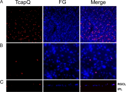
Fig. 5.
Intracellular TcapQ activation in retrogradely labeled retinal ganglion cells. RGCs were retrogradely labeled by injection of FluoroGold (FG) 7 days before induction of RGC apoptosis. Retinal flatmounts were prepared after 6 h of exposure to 25 nmol NMDA and subsequent TcapQ injection (A and B). Vertical retinal sections were then prepared from the same retinae (C). Intracellular probe activation (red) and FG labeled RGCs (blue) are confirmed to be colocalized (pink) in merged overlay images from retinal flatmounts at low (A) and high (B) magnification. (C) Vertical retinal sections confirm probe activation in retrogradely labeled RGCs in the RGC layer (RGCL). IPL, inner plexiform layer. (Magnification: A and C, ×10; B, ×40.)

