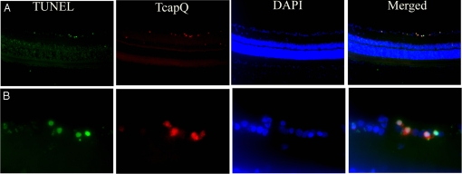
Fig. 6.
TcapQ activation corresponding to retinal ganglion cell apoptosis in the NMDA model. TUNEL assay was performed on frozen vertical retinal sections from eyes exposed to 80 nmol NMDA with subsequent intravitreal injection of TcapQ. Retinas were prepared and examined by fluorescence microscopy to identify apoptotic TUNEL labeled cells (green), TcapQ activated cells (red), and DAPI stained cell nuclei (blue). As with TUNEL labeling, TcapQ activation was primarily limited to cell bodies in the RGC layer consistent with RGCs. DAPI staining reveals the retinal architecture. Merged images confirm that TcapQ activation colabels TUNEL-positive cells. (Magnification: A, ×10; B, ×40.)

