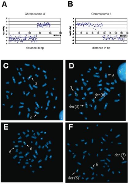
Figure 1.
Panels A and B: Array Painting analysis demonstrating changes in hybridization signals at translocation breakpoints on chromosomes 3 and 6. Panels C–F: metaphase spreads prepared from lymphoblast cell line established from normal control (C and E) and patient with t(3;6) translocation (D and F). C and D show chromosome 3 identified using chromosome 3 specific centromeric probe, labeled with spectrum-orange. Normal and t(3;6) spreads were hybridized with a spectrum-green labeled chromosome 3 BAC probe, RP11-354M3, that contained the breakpoint (as shown by the split signal). E and F show corresponding images for chromosome 6. The breakpoint mapped within RP11-44P5 as shown by the split signal. [Color figure can be viewed in the online issue, which is available at www.interscience.wiley.com]

