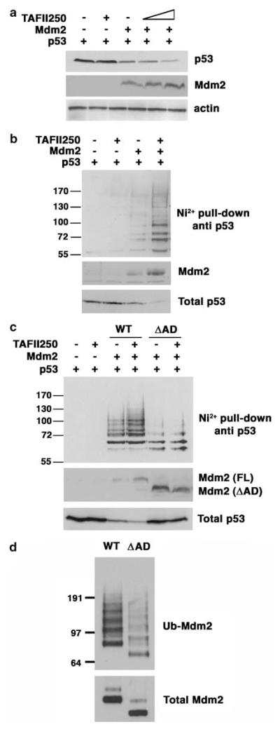
Figure 3.
TAFII250 promotes ubiquitylation and degradation of p53. (a) H1299 cells were cotransfected with 1 μg of p53, 5 μg of Mdm2 and 5 and 10 μg of TAFII250 expression vectors. Cells were harvested 36 h after transfection and lysed with 2 × SDS sample buffer. The cells extracts were analysed for p53 protein levels. (b) Ni2+ pull-down was performed using extracts of H1299 cells that had been transfected with 1 μg of p53, 1 μg of Mdm2, 1 μg of TAFII250 and 2 μg of His-Ubiquitin expression vectors. The His-tagged ubiquitylated proteins were purified with Ni2+ agarose beads and analysed by western blotting using anti-p53 antibody DO-1. (c) p53 ubiquitylation was measured in H1299 cells transfected with plasmids encoding p53, TAFII250 and Mdm2 (wild-type and ΔAD mutant) as indicated. (d) Mdm2 (wild-type and ΔAD mutant) auto-ubiquitylation was measured in H1299 cells transfected with plasmids encoding wild type or ΔAD mutant Mdm2.

