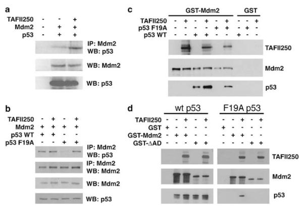
Figure 6.
TAFII250 promotes the interaction of Mdm2 and p53. (a) H1299 cells were transfected with plasmids expressing Mdm2 alone or Mdm2 together with TAFII250. Cell extracts were prepared and the volumes adjusted empirically to yield equal concentrations of Mdm2 in each extract. H1299 cells were also transfected, separately, with a plasmid expressing wild-type human p53. Subsequently, extracts were prepared and equal volumes mixed with the extracts from the Mdm2- or Mdm2/TAFII250-expressing cells. Mdm2 was subsequently immunoprecipitated and the amount of co-immunoprecipitating p53 was measured by Western blotting using the anti-p53 polyclonal antibody, CM-1. (b) H1299 cells were transfected with plasmids encoding TAFII250, Mdm2 and wild type or a F19A mutant of p53. Cells were treated with the proteasome inhibitor, MG132, 6 h before harvesting. Mdm2 and p53 were detected by Western blotting following immunoprecipitation of Mdm2 (upper panels). The total levels of Mdm2 and p53 are also shown (lower panels). (c and d) GST-Mdm2, GST-Mdm2-ΔAD (d) or GST alone were bound to glutathione-sepharose beads and subsequently incubated with p53 WT or a F19A mutant of p53 in the presence or absence of TAFII250. Immobilized proteins were detected by Western blotting.

