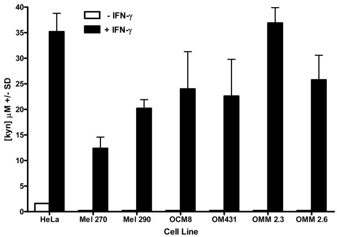
Figure 4. IDO expressed by IFN-γ-stimulated primary and metastatic uveal melanoma degrade tryptophan and produce kynurenine.
IDO activity by uveal melanoma was assessed by colorimetric detection of kynurenine in uveal melanoma culture supernatants using a modified assay by Kudo and Boyd (2000). Uveal melanoma cells were cultured in either 2.5uM tryptophan medium, with or without IFN-γ for 72 hours. Supernatants were collected, treated with TCA to precipitate proteins, and mixed with p-dimethylaminobenzaldehyde to stabilize kynurenine in the samples. Kynurenine was detected by colorimetric analysis using a spectrophotometer at a wavelength of A490. Kynurenine concentration in uveal melanoma culture supernatants was determined by standard regression analysis derived from colorimetric analysis of kynurenine concentration standards. The results are representative of 4 separate experiments.

