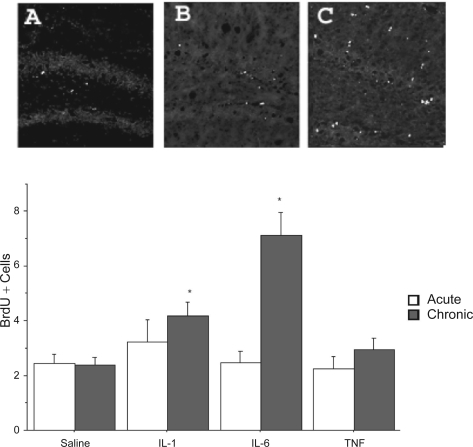
Figure 3.
The photomicrographs depict BrdU-positive cells within the dentate gyrus following a single or repeated intra-hippocampal cytokine or saline infusion. Elevated BrdU staining within the dentate gyrus was observed in animals subjected to repeated intra-hippocampal infusion of either IL-1β (panel B) or IL-6 (panel C), relative to animals that received vehicle (panel A). The BrdU increase was particularly robust for the repeated IL-6 treatment. Quantification of mean (± SEM) number of BrdU-positive cells per section confirmed that repeated (grey bars) but not a single (white bars) intra-hippocampal infusion of IL-1β and IL-6 significantly increased BrdU staining, relative to animals that received infusion of saline.
Notes: *p < 0.05 vs saline-treated animals, 10× magnification.

