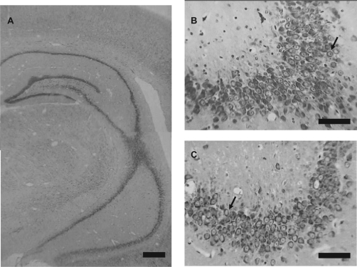
Figure 5.
Photomicrographs of coronal section through the brains of animals which had neonatal bilateral administration of lidocaine into the ventral hippocampus (A and C) and control animals (B). Panel A shows that lidocaine-treated animals had the dorsal and ventral portion of hippocampus morphologically intact without gliosis. When we amplified the CA3 hippocampal region (B and C) we observed that control animals (B) had normal morphology represented by neurons with distinguishable nucleus, nucleolus, and a high ratio of nucleus/cytoplasm (arrows in B demonstrated normal neuron of this region), whereas neonatal bilateral administration of lidocaine into the ventral hippocampus caused a mild reduction of neurons with some alterations such as chromatin condensation, nucleolus loss, and cell shrinkage, but no observed glial proliferation (arrows in C demonstrated alterations).
Note: Bar in A represents 250 μm and bars in B and C represent 50 μm.

