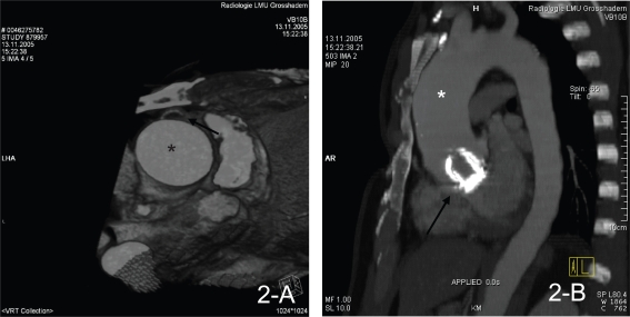
Figure 2.
ECG-gated 64-row MSCT with volume rendering technique (VRT) and three-dimensional reconstruction demonstrating the aneurysmatic ascending aorta (*, 2-A, -B), the sternum proximity to the RCA (arrow, 2-A) as well as the prosthetic aortic valve (arrow, 2-B).
Abbreviations: ECG, electrocardiogram; MSCT, multi-slice computed tomography; RCA, right coronary artery.

