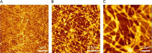
Figure 2.
AFM images of RADA16-I. The working solution of RADA16-I was prepared at 100 μM concentration by using Milli-Q water. The nanostructure of RADA16-I was scanned at different scales, 5 × 5 μm2 (A), 2 × 2 μm2 (B) and 1 × 1 μm2 (C). Dense nanofibers were observed, indicating peptide RADA16-I has excellent self-assembling ability to form nanofiber scaffolds.

