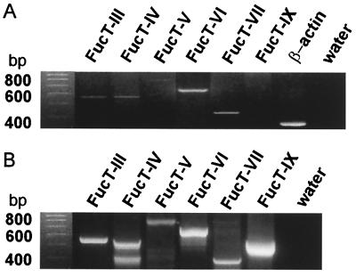
Figure 1.
Detection of FucTs by RT-PCR. RT-PCR products separated on 3% NuSieve agarose gel and stained with ethidium bromide are shown (A). Positive-control reactions for the FucT-PCR were performed on genomic DNA and are represented in B. RNA quality was checked with the amplification of β-actin (A). The H2O controls are shown on the rightmost lane. A 100-bp ladder was used as standard (first lane).

