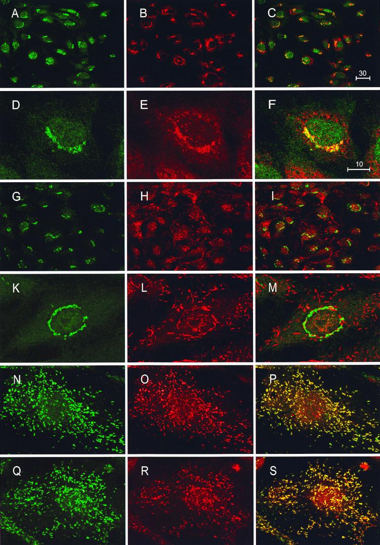
Figure 2.
Immunolocalization of FucT-VI in HUVECs. Dual confocal immunofluorescence on HUVECs was performed as described in Materials and Methods. A double staining for GalT-1 (A and D) and FucT-VI/III (B and E) with the OLI antibody is shown in an overview in the first row (A–C) and on a single cell level in the second row (D–F). The overlays of the respective pictures are given in the last column. Colocalization resulted in a yellow color. Bars = 30 μm and 10 μm, respectively. GalT-1/PEP6B (G, H, K, and L) dual staining is represented in rows 3 (overview) and 4 (single cell). Single-cell sections of double stainings with P-selectin (N)/PEP6B and vWF (Q)/PEP6B are shown in row 5 (N–P) and 6 (Q–S).

