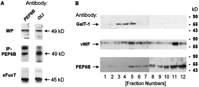
Figure 3.
Immunodetection of FucT-VI in human endothelial cells. HUVECs were analyzed by SDS/PAGE followed by immunoblotting for the presence of FucT-VI (A). In the WP bodies enriched fraction (WP), both antibodies PEP6B (lane 1) and OLI (lane 2) recognized a major band migrating at 49 kDa (A, row 1). A band of the same molecular weight was obtained after immunoprecipitation with PEP6B from HUVEC lysate given in row 2 (IP-PEP6B). Immunostaining of recombinant soluble FucT-VI (sFucT) is shown as a control (bottom row). In B, the localization of FucT-VI by PEP6B in Percoll density gradient fractions (see Materials and Methods) is represented. Fractions were tested for the presence of GalT-1 (row 1), vWF (row 2), and FucT-VI with the PEP6B antibody (row 3). The molecular standards are indicated (Right).

