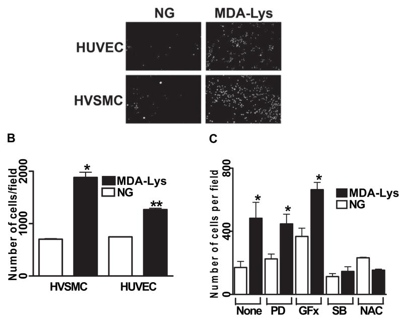
FIG. 7.
A: MDA-Lys treatment increases monocytes adhesion to HVSMC and HUVECs. THP-1 cells were cultured with or without MDA-Lys for 12 h and then labeled with fluorescent dye Calcein-AM for 15 min at 37°C. Labeled THP-1 cells were allowed to adhere to either HUVEC or HVSMC monolayers in 24-well culture dishes. After careful washing, specifically bound monocytes were counted as described previously (19). Results are expressed as number of monocytes bound per high-power field. B: Bar graph shows means ± SE from three to five experiments (*P < 0.005, **P < 0.01 vs. respective controls). C: THP-1 cells were treated for 12 h with the indicated inhibitors or corresponding vehicle and then treated with or without MDA-Lys and then binding to HVSMC examined as above. Results shown are means ± SE (n = 4; *P < 0.01 vs. respective control). PD, 2′-amino-3′methoxyflavone (PD-98059); SB, 4-[4-(4-fluorophenyl)-5-(4-pyridinyl)-1H-imidazol-2-yl]phenol (SB202190).

