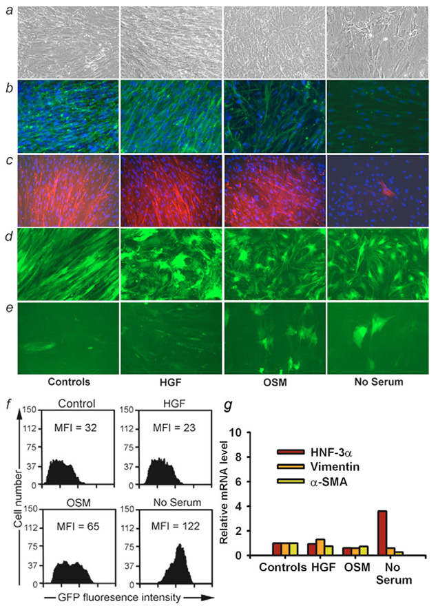
Fig. 8.
Regulation of meso-endodermal phenotype. Perturbations in P3 fetal liver cells cultured with FBS (control) or hHGF, OSM and no serum. (a) Cell morphology under phase contrast with more compact cells in OSM or no serum. (b) Vimentin staining (green color). (c) α-SMA staining (red color) with characteristic filamentous pattern seen in mesenchymal cells and less pronounced expression in cells cultured with no serum (panel on extreme right). (d,e) Cells transduced with GFP lentiviral vector under PGK or Alb promoter, respectively. While PGK expression remained unaltered, Alb promoter was more active in cells cultured with OSM or no serum. Magnification: 200×. (f) Flow cytometry analysis indicating twofold and fourfold greater mean fluorescence intensity (MFI) of GFP expression under Alb promoter in cells cultured with OSM or no serum. (g) Changes in gene expression in P3 cells with qRT-PCR using β2-microglobulin as internal control. Vimentin expression declined to 60% of controls and α-SMA expression declined to 72% and 26% of controls in cells cultured with OSM or no serum, respectively. In cells without serum, HNF-3α (FOXA1) expression increased.

