Fig. 9.
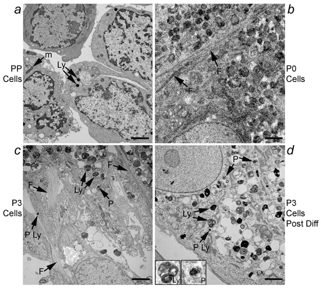
EM analysis of cellular ultrastructure. (a) Freshly isolated PP cells with large nuclei, prominent nucleoli and relatively sparse cytoplasm with scattered mitochondria (m) and secondary lysosomes (Ly). P0 cells (b) and P3 cells (c) that had been in culture for three weeks and seven weeks, respectively. These cells showed intermediate filaments (F, arrows) as well as complex cytoplasmic appearance with multiple mitochondria, vacuoles, occasional primary lysosomes (P Ly) as well as secondary lysosomes and microperoxisomes (P) consistent with alterations in cell morphology and presence of both mesenchymal and epithelial features. (d) P3 cells cultured without serum. Under this condition, most cells no longer contained intermediate filaments, although the epithelial phenotype did not resemble that of freshly isolated PP cells, indicating the incompleteness of phenotypic reversion. These cells again contained primary and secondary lysosomes as well as microperoxisomes, which are additionally shown in insets. Scale bars: 2 µm.

