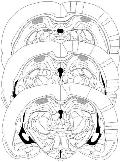
Figure 2. Cannulae Placements and Drug Infusions.
Schematic representations of rat brain sections at three rostrocaudal planes (−3.80, −4.30, and −4.80 from bregma) taken from the atlas of Paxinos and Watson, showing, in stippling, the extension of the area reached by the infusions in the dorsal hippocampus. Reprinted from The Rat Brain in Stereotaxic Coordinates by Paxinos and Watson, pages 33, 35, and 37, Academic Press (1997), with permission from Elsevier.

