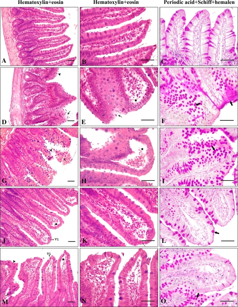
Fig. 1.
Histological examination of rat small intestine taken from the segments of ileum. Photomicrographs of ileum sections stained with H + E (a, b, d, e, g, h, j, k, m, n), and PAS + Hl (c, f, i, l, o). a–c Control rats ileum; showing normal morphology. d–f Single dose radiation-treated rats; shortened and thickened villi (arrowhead) and degenerative changes in the epithelial cells (arrow), lifting of epithelial layer from the lamma propria (asterisks), and increase in the goblet cells and mucins showing strongly positive to PAS staining (thick arrows). g–i Two dose radiation-treated rats; shortened and irregular villi (arrowhead) and breaking in the epithelial cells (arrow), massive subepithelial lifting and capillary congestion in the villus (asterisks), and increase in the goblet cells and mucins showing strongly positive to PAS staining (thick arrows). j–l Single dose radiation-treated with curcumin rats; villi were generally normal (Vi) although apical regions of some villi were lightly subepithelial lifting (asterisks) and thinning (arrow), normal scattered goblet cells (thick arrows). m–o Two dose radiation-treated with curcumin rats; development of subepithelial space (asterisks) and lightly capillary congestion (thick arrows) usually at the apex of the villus, occasional occurrence of thickened villi (arrowhead) along with normal villi (Vi) and breaking in the epithelial cells (arrow), goblet cells showing strongly positive to PAS staining (thick arrows) (scale bar: 50 μm)

