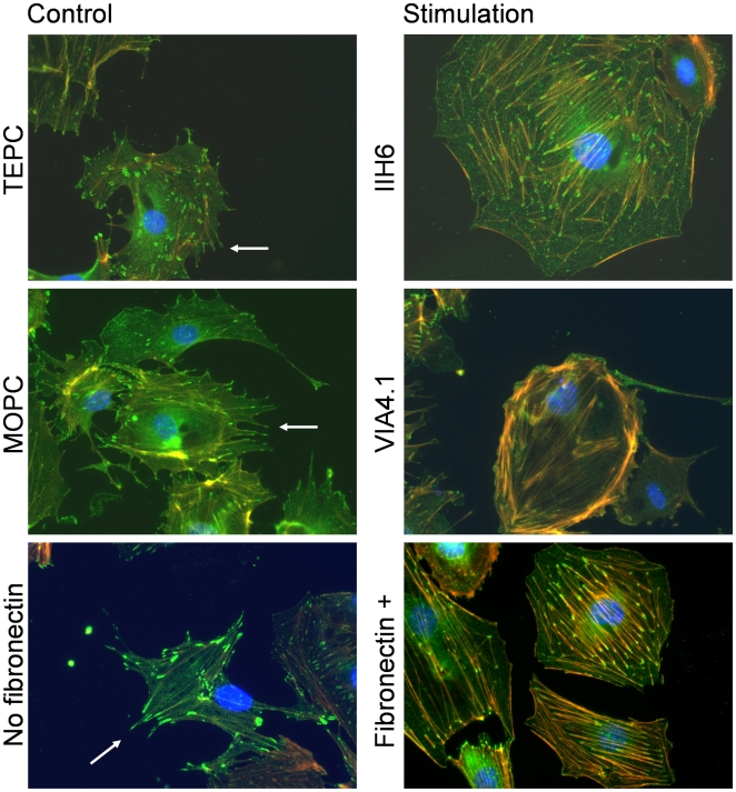
Figure 4. Ligation of α-dystroglycan results in a loss of pedicles in cultured mouse podocytes.
Differentiated podocytes on collagen coatings were incubated with either monoclonal antibodies IIH6 or VIA 4.1, their respective isotype controls (TEPC or MOPC) or fibronectin. The podocyte pedicles (arrow) are probed with antibodies against Mena (green, which also localizes along stress fibers and in focal contacts at the tips of stress fibers [58], [65]). The pedicles were lost after 6 hours of incubation with the antibodies. As these pedicles may be the in vitro manifestation of foot processes, their retraction may imply foot process effacement. Percentage of podocytes with smooth surfaces were counted, which yielded for IIH6 60% versus TEPC 30% (χ2 = 20.8, p<0.001), for VIA4.1 51% versus MOPC 28% (χ2 = 9.01, p<0.01) and fibronectin 38% versus control 30% (χ2 = 1.3, p = 0.25). (Red = phalloidin (actin); blue = DAPI (DNA)).

