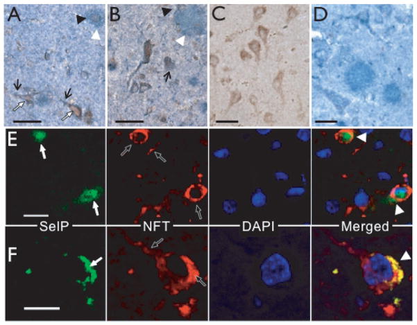
Fig. 4.
Association of SelP with NFT immunoreactivity. A,B: NFT-immunopositive pyramidal cells with apical dendrites showing abnormal architecture (white arrows) also expressed SelP. The NFT antibody also labeled dystrophic neurites (black arrows) that were associated with SelP-positive cells (black arrowheads). C: Negative control for double labeling with NFT antibody omitted. D: Negative control for double labeling with SelP antibody omitted. E–F: SelP (green, white arrows) and NFT immunoreactivity (red, black arrows), with DAPI-stained nuclei (blue). Merged images on lower right show yellow co-localization of SelP with NFTs (white arrowheads). Scale bars: A–E, 20 μm; C, 10 μm.

