
Figure 10.
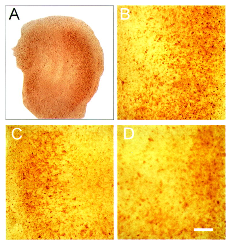
Bright-field photomicrograph of proliferating cell nuclear antigen (PCNA) immunostaining 48 h after 10 μM N-methyl-D-aspartate (NMDA) exposure in hippocampal organotypic cultures. Low-power image (A) has the same orientation as previous figures. NMDA exposure caused an increase in PCNA-positive cells in CA1 (B), CA3 (C), and the dentate (D). Bar = 50 μm.