
Figure 56.
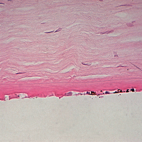
Fuchs corneal dystrophy. Light microscopic appearance of the corneal endothelium, Descemet membrane, and the adjacent corneal stroma showing guttae, a paucity of endothelial cells including some containing melanosomes. Hematoxylin and eosin stain.