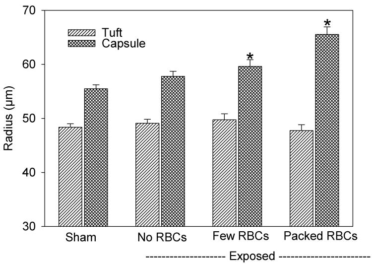
Figure 3.
A plot of measurements of the radii of the glomerular tuft and capsule in histological samples taken 5 min after exposure. For glomeruli with GCH, the radius of the capsule was significantly (*) increased (P<0.05), but the radius of the tuft was unchanged, relative to shams. The width of Bowman’s space (the capsule radius minus the tuft radius) was approximately doubled for glomeruli with GCH.

