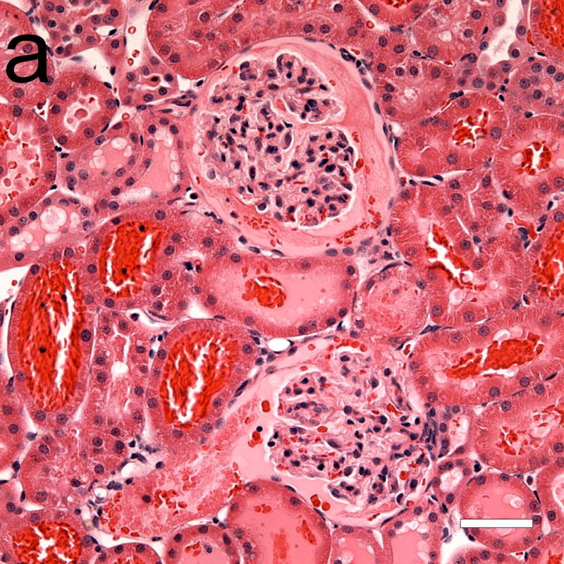
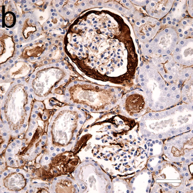
Figure 4.


Photomicrographs of adjacent serial sections (a) from a 4 h sample stained with hematoxylin and eosin, and (b) with hematoxylin and labeled anti-fibrinogen antibody (dark frown stain). Some light staining of glomerular and peritubular capillaries was normal, and was also seen in shams. Although the two glomeruli in (a) seem mostly free of red cells from GCH, the anti-fibrinogen stain reveals large areas occupied by fibrin clots (b). Scale bars: 50 μm.
