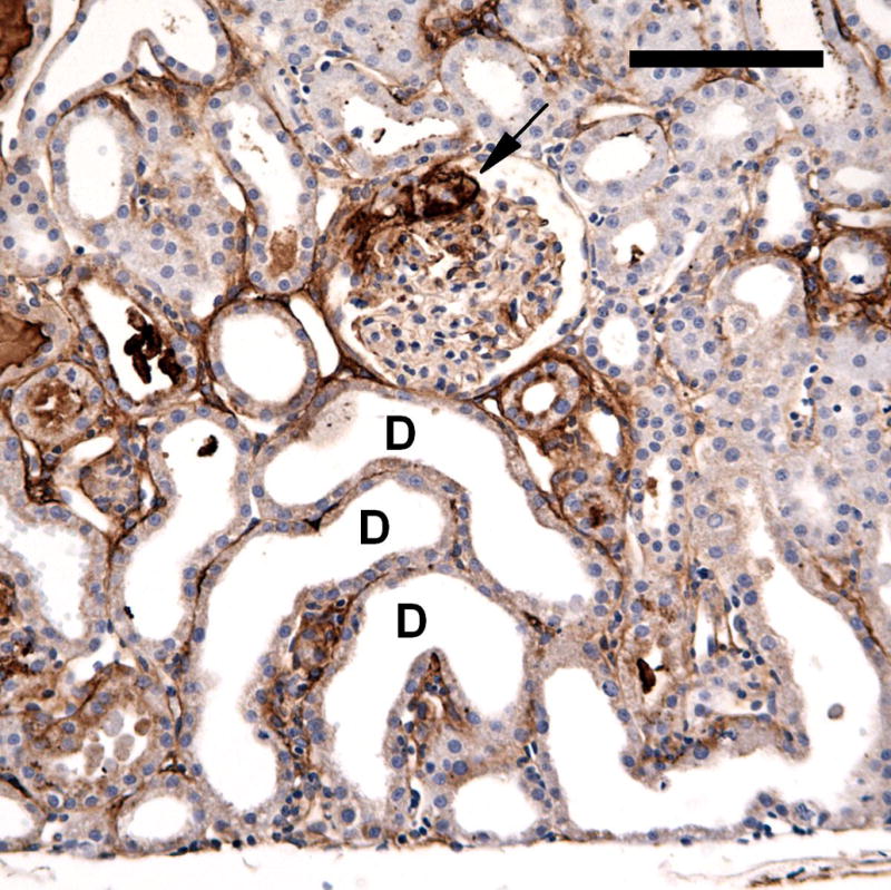
Figure 5.

A photomicrograph of an anti-fibrinogen stained section from a 2 day post-exposure sample. A glomerulus retains some fibrin clot (arrow) and some nearby tubules have become dilatated (some examples marked D). The tubular dilatation represented tubular injury, with swelling and loss of the brush border of the epithelial cells. Scale bar: 100 μm.
