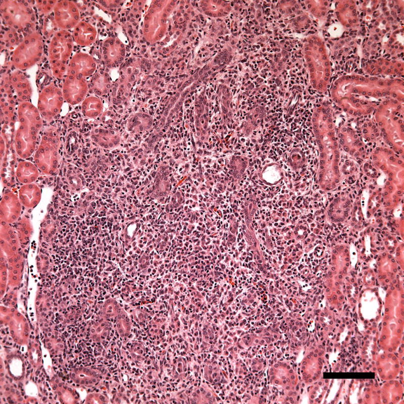
Figure 7.

A photomicrograph of a histological sample taken 1 week post-exposure showing a region with inflammatory cell infiltration (probably neutrophils). The inflammatory response may represent removal of necrotic debris during healing. Scale Bar: 100 μm.
