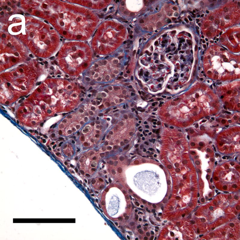
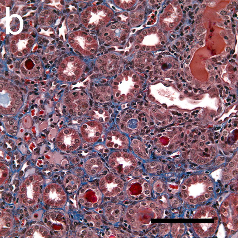
Figure 8.


Photomicrographs of histological sections taken 4 weeks after exposure which were stained with Masson’s trichrome stain to show collagen (blue). The collagen formation around some tubules in the cortex (a) indicated a progression of interstitial fibrosis in response to the tubular injury following GCH. Some tubules in the medulla (b) also appeared to have retained material from the initial GCH, with the lumen of the tubules appearing red or pink (rather than clear white). Scale Bar: 100 μm.
