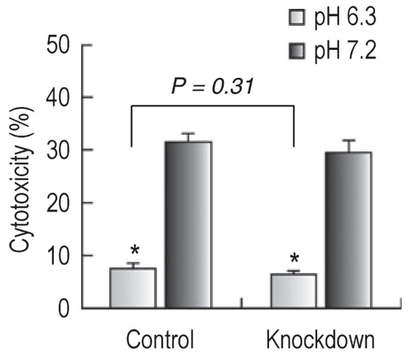
Fig. 6.

Glutamate cytotoxicity in neurons at acidic and normal pH. Neurons with siRNA control and NBCn1 knockdown lentivirus (72 h post-infection) were exposed to 500 μM glutamate (50 μM glycine and 0 Mg2+, pH 7.2) for 10 min and then switched to medium at pH 6.3 or 7.2 for 6 h without the added glutamate. Cytotoxicity was determined as the percentage of LDH released from neurons relative to the total release after background subtraction (n = 5 for each groups of neurons). Error bars are SEM. *P < 0.05.
