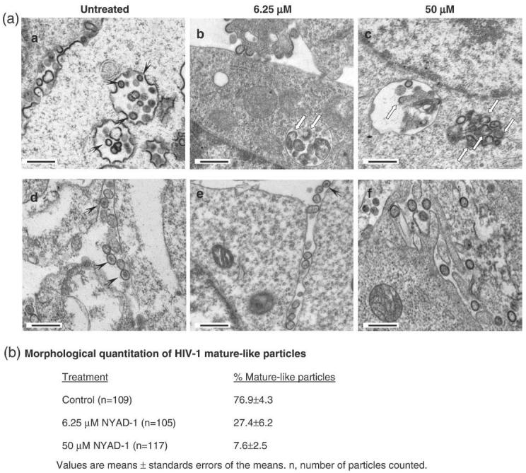
Fig. 8.

Electron microscopic analysis of HIV-1 virus-like particles produced in the presence of 6.25 μM and 50 μM NYAD-1. (a) 293T cells expressing Gag (upper panel) or Gag-Pol(lower panel) were incubated with 2 ml of culture medium containing no peptide or 6.25 μMor50 μM NYAD-1 4 h post transfection with vectors encoding Gag or Gag-Pol. At 24 h post transfection, cells were processed and examined under the electron microscope. The scale bar represents 500 nm; (b) the morphological quantification of mature-like particles.
