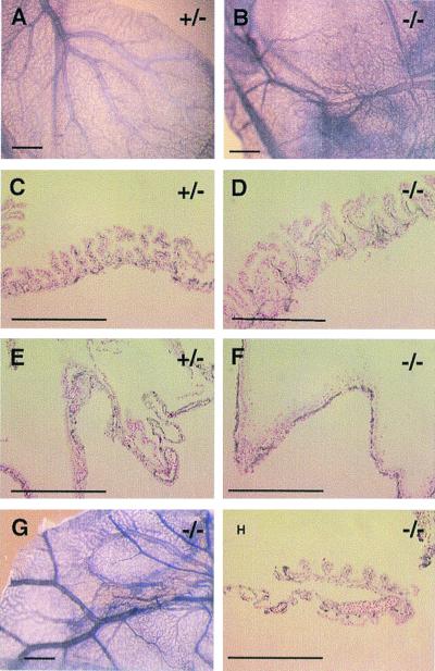
Figure 3.
Immunohistochemical staining of normal and mutant yolk sac membranes (all at E11.5) with anti-CD31 antibody. (A) Normal yolk sac membrane, showing a highly organized vascular tree pattern. (B) The vascular pattern from a severely hemorrhaging mutant differs from the normal control only slightly when viewed at the whole-mount level. (C–F) Histological sections of anti-CD31-stained specimens. (D and F) In most severe mutants, about a third of endothelial cells form extended endothelial sheets without proper lumen structures. (G and H) Moderately hemorrhaging mutants had no significant vascular disorganization, when viewed both at the whole-mount level and in histological sections. (Bars: A, B, and G, 600 μm; C–F and H,100 μm.)

