
Fig. 2.
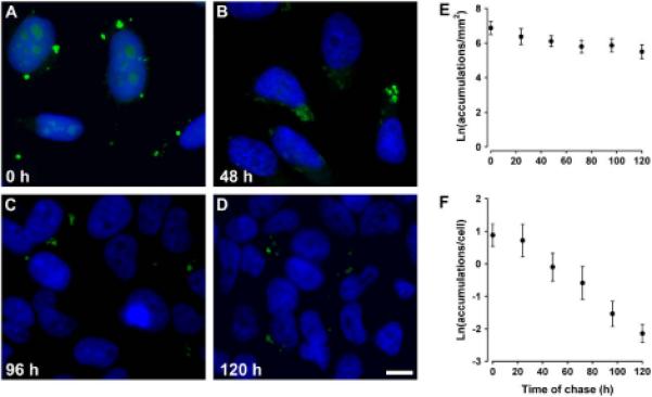
CX50P88S accumulations are long-lived. (A-D) Photomicrographs of HeLa-CX50P88S(Cys)4 cells that were pulse-labeled with FlAsH, chased in normal growth medium for 0 (A), 48 (B), 96 (C) or 120 (D) h, fixed, and stained with DAPI to visualize cell nuclei. (E, F) Semi-logarithmic graphs show the quantification of the number of FlAsH-labeled CX50P88S accumulations per mm2 (E) or per number of cells (F) as a function of the time of chase in hours. Scale bar, 20 μm.