
Fig. 6.
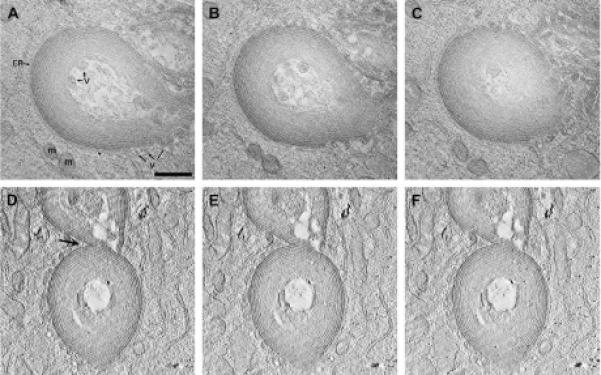
Three-dimensional reconstructions of multilamellar accumulations. Thick sections of photoconverted CX50P88S(Cys)4 accumulations were prepared for electron tomography. Two tomograms were reconstructed and analyzed. (A-C) Slices from the top, middle and bottom parts of the double tilt reconstruction are shown to illustrate the “onion skin” appearance of these structures. Also indicated in the slice shown in (A) are the close apposition of ER membranes (ER), mitochondria (m) and vesicles (v). (D-F) On occasion, two accumulations were joined together by a piece of ER attached to both (see arrow), as seen in three slices from this single tilt tomogram. Scale bar, 0.5 μm.