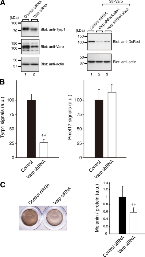
Figure 6.
Reduced expression of Tyrp1 in Varp-deficient melanocytes. (A) Reduced expression of Tyrp1 and Varp in siVarp-treated melanocytes as revealed by immunoblotting (left panel). Cell lysates of melan-a cells treated with either siVarp or control siRNA (∼20 μg) were subjected to 10% SDS-polyacrylamide gel electrophoresis (PAGE) followed by immunoblotting with anti-Varp specific antibody (1/100 dilution), anti-Tyrp1 antibody (1/200 dilution), and anti-actin antibody (1/400 dilution). Right, efficiency and specificity of shRNA targeted against Varp were shown. Str-Varp was expressed in COS-7 cells together with Varp shRNA or a control vector. Cell lysates were subjected to 10% SDS-PAGE followed by immunoblotting with anti-DsRed antibody (1/100 dilution) and anti-actin antibody. The positions of the molecular mass markers (× 10−3) are shown on the left. (B) Reduced expression of Tyrp1 in Varp shRNA-expressing melanocytes as revealed by immunofluorescence analysis. Note that expression of Tyrp1 in Varp-deficient cells dramatically reduced, whereas expression of Pmel17 had no effect. The bars represent the means ± SE of data from three independent dishes (n > 50). **p < 0.01, Student's unpaired t test. (C) Reduced melanin content in Varp siRNA-expressing melanocytes. Melan-a cells expressing Varp siRNA (right) and control siRNA (left) were harvested, and their melanin content was assayed by measuring optical density at 490 nm. The bars represent the means ± SD of data from triplicate experiments. **p < 0.01, Student's unpaired t test.

