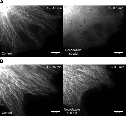
Figure 6.
Microtubule deformation in the absence of polymerization. Distribution of microtubules in the periphery of GFP-tubulin–labeled LLC-PK1 epithelial cells is shown before and after exposure to different concentrations of the drug nocodazole. (A) Nocodazole is used at 50 μM. (B) Nocodazole is used at 100 nM. Time (minutes) is in relation to the addition of nocodazole. Horizontal bar, 5 μm.

