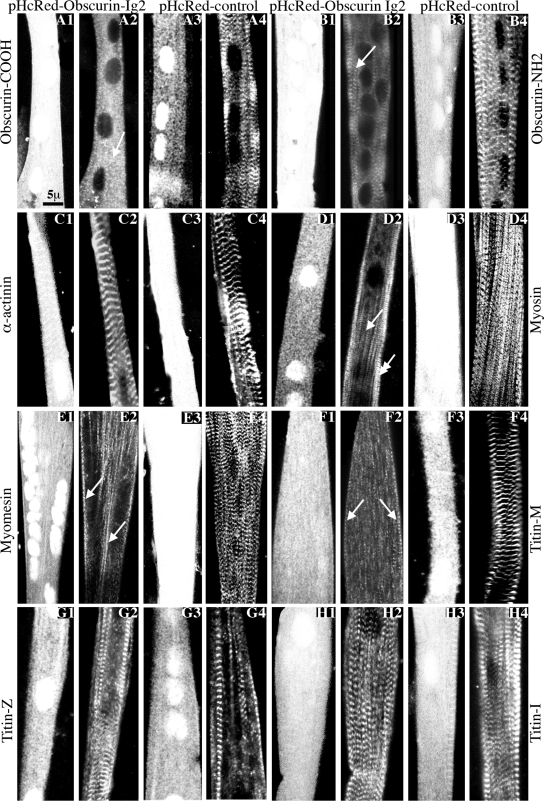
Figure 1.
Adenoviral overexpression of the Ig2 domain of obscurin in primary cultures of rat skeletal myotubes resulted in disorganized M- and A-bands but not Z-disks or I-bands. (A1–A4 and B1–B4) The striated distribution of endogenous obscurin was severely altered after overexpression of pHcRed-Obscurin-Ig2 virus (A1 and A2 and B1 and B2), but not control pHcRed virus (A3 and A4 and B3 and B4) as shown by immunostaining with antibodies to the COOH terminus (A2, arrow; and A4) or the NH2 terminus (B2, arrow; and B4) of obscurin (odd numbered panels show pHcRed fluorescence). (C1–C4) The regular organization of α-actinin at Z-disks was unaffected after overexpression of pHcRed-Obscurin-Ig2 (C1 and C2) or control pHcRed (C3 and C4) virus. (D1–D4) Sarcomeric myosin exhibited adiffuse cytoplasmic distribution (single arrow) with residual accumulation in striated structures at the cell periphery (double arrow) in cultures infected with pHcRed-Obscurin-Ig2 virus (D1-D2) but not in cultures infected with pHcRed virus where it assumed its typical periodic organization at A-bands (D3 and D4). (E1-E4 and F1-F4) Similar to sarcomeric myosin, myomesin (E1 and E2, arrows) and COOH-terminal epitopes of titin present at the M-band (F1 and F2, arrows) were primarily detected along fibrils showing occasional periodicity after overexpression of pHcRed-Obscurin-Ig2 but not control virus (E3 and E4 and F3 and F4, respectively). (G1-G4 and H1-H4) Strong periodic labeling of the NH2 terminus (G1-G4) and middle portion (H1-H4) of titin at the Z-disk and I-band, respectively, was observed in myotubes infected with either pHcRed-Obscurin-Ig2 (G1 and G2 and H1 and H2) or control pHcRed virus (G3 and G4 and H3 and H4).

