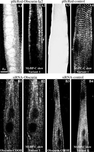
Figure 8.
MyBP-C slow variant-1 failed to assemble into M-bands in primary cultures of skeletal myotubes after manipulation of the expression of obscurin. (A1–A4) Confocal images of P1 myotubes treated with pHcRed-Obscurin-Ig2 virus (A1) and stained with antibodies specific to MyBP-C slow variant-1 (A2); MyBP-C slow variant-1 failed to organize at M-bands, after overexpression of the Ig2 repeat of obscurin and concentrated along fibrillar structures showing occasional periodicity (A2, arrow). This was not the case in cells infected with control pHcRed virus (A3) in which MyBP-C slow variant-1 showed a regular distribution at M-bands. (B1–B4) Confocal images of P1 myotubes treated with a siRNA virus that specifically targets obscurin and labeled with antibodies to the COOH terminus of obscurin (B1) and antibodies specific for MyBP-C slow variant-1 (B2); MyBP-C slow variant-1 failed to assemble into M-bands when obscurin was knocked down and concentrated in the same structures as residual obscurin did (arrows). In cells infected with control siRNA virus, both obscurin (B3) and MyBP-C slow variant-1 (B4) assumed their typical organization at M-bands.

