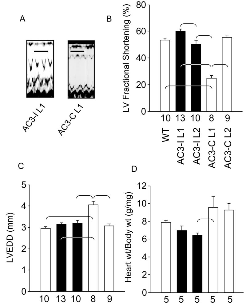
Figure 2. AC3-I transgenic mice have preserved LV size and shortening.
a. Representative M-mode echocardiography tracings from AC3-I L1 and AC3-C L1 mice. Calibration bars indicate 200 ms. b. Summary data of left ventricular (LV) fractional shortening. Fractional shortening was significantly different between lines (P<0.001). The brackets indicate significant (P<0.05) post-hoc comparison differences in panels b–d. c. Summary data for LV end-diastolic diameter (LVEDD) measurements. LVEDD was significantly different between lines (P<0.001). d. Heart weight (wt) to body wt ratios were significantly different between groups (P=0.012). The number of animals studied is indicated by numerals on the abscissa.

