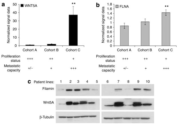
Figure 1. Filamin A expression is increased in primary melanoma cell lines with high Wnt5A expression.
(a) WNT5A and (b) FLNA mRNA expression in the three cohorts of the Mannheim data set. Cohorts A (n=19) and B (n=10) were considered low metastatic cell lines, whereas cohort C (n=16) was highly metastatic. Their metastasis rates inversely correlate with their proliferation as indicated in the table below the graphs. FLNA was significantly correlated with WNT5A (**P<0.01). Bars represent averages of all patients in each cohort. (c) Western blot analysis was performed on 10 melanoma patient cell lines to detect filamin A and Wnt5A, and confirmed that filamin A protein expression and Wnt5A expression are correlated at the protein level as well. β-Tubulin was used as a loading control.

