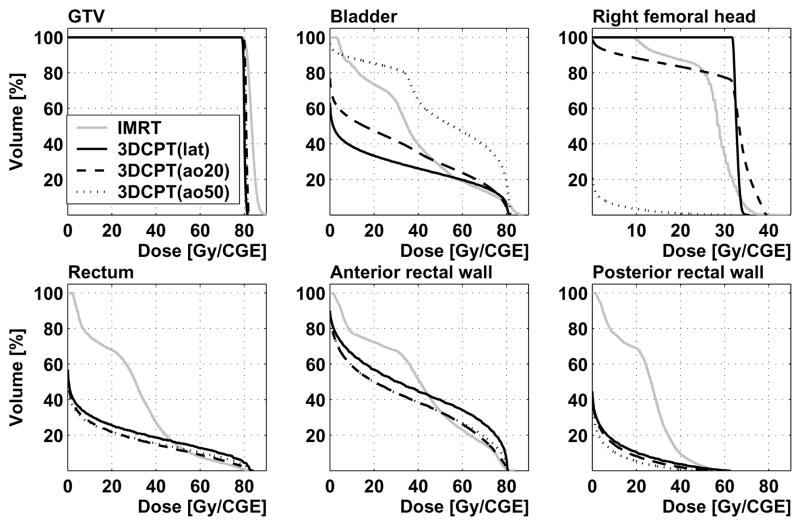
Figure 5.
Patient 2: dose-volume histograms from IMRT and 3D-CPT plans with lateral parallel-opposed (“lat”), and anterior-oblique configurations with the beams rotated by 20° (“ao20”) and 50° (“ao50”) towards the anterior. DVH are shown for the gross tumor volume, bladder, right femoral head, whole rectum, anterior and posterior rectal walls.

